Single Chamber Pacemaker on Chest X-ray PA
Single chamber pacemaker on Chest X-ray PA
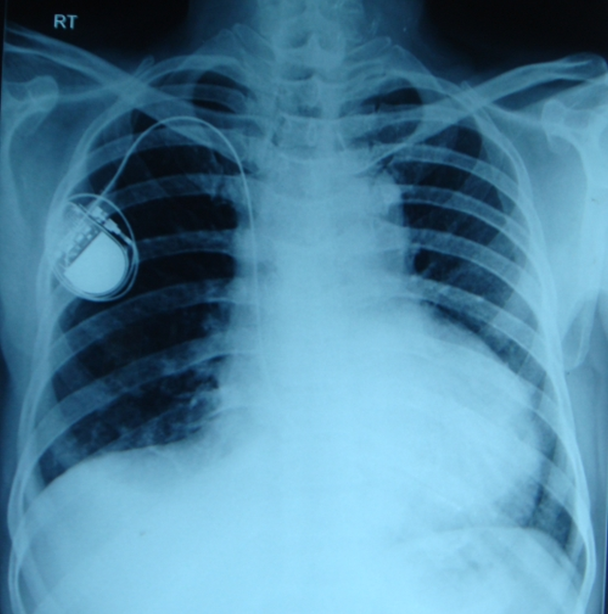
The device seen in the upper portion of the right hemithorax is a single chamber pacemaker. The lead can be seen screwed into the device and lead courses out the pacemaker pocket with a loop going beneath the right clavicle. Further course along the right cardiac border is through the superior vena cava into the the right atrium. The lead enters the circulation through the subclavian vein between the clavicle and first rib. This is a region where it can be damaged by “subclavian crush”.
From the right atrium the lead crosses the midline to enter the right ventricle and the tip is at the right ventricular apex. That portion of the lead will be seen well only in penetrated views or on fluoroscopy. In dual chamber pacemakers, two leads can be seen with one lead in the atrium and another in the ventricle. Three chamber pacing is done in cardiac resynchronization therapy where one lead each is seen in right atrium, right ventricle and left ventricle. Left ventricular lead is placed within a coronary vein, after passing through the coronary sinus, usually on the lateral wall of the left ventricle. While the right atrial and right ventricular leads pace endocardially, the left ventricular lead paces epicardially.
Fluoroscopic imaging of the pacemaker and lead is an integral part of pacing system malfunction evaluation. Lead fractures and dislodgements can be identified by fluoroscopy using an image intensifier. Occasionally, too many loops of the lead may be seen within the pacemaker pocket in pacemaker twiddler’s syndrome, in which the recipient rotates the pacemaker within its pocket, bringing the lead out of the heart. This results in loss of capture.
Related Posts


About The Author
Johnson Francis
Former Professor of Cardiology, Calicut Govt. Medical Kozhikode, Kerala, India. Editor-in-Chief, BMH Medical Journal