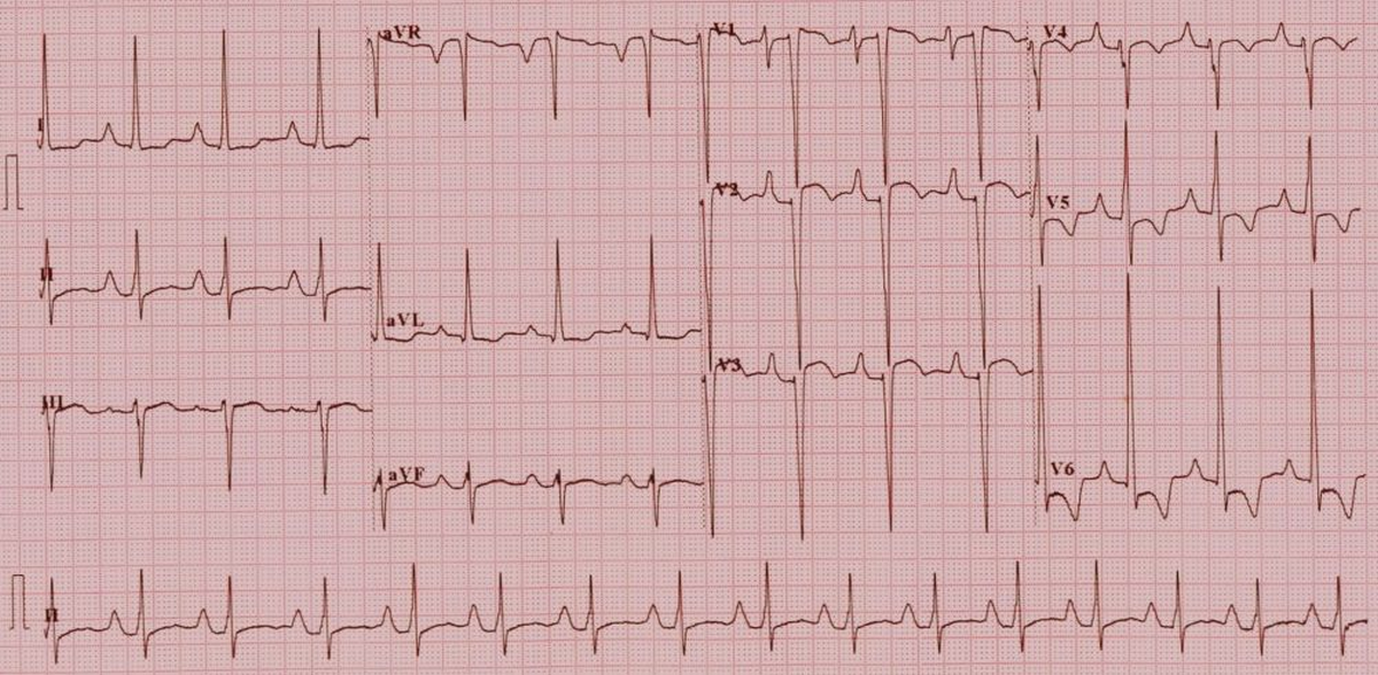
Tricuspid atresia – ECG
Tricuspid atresia – ECG
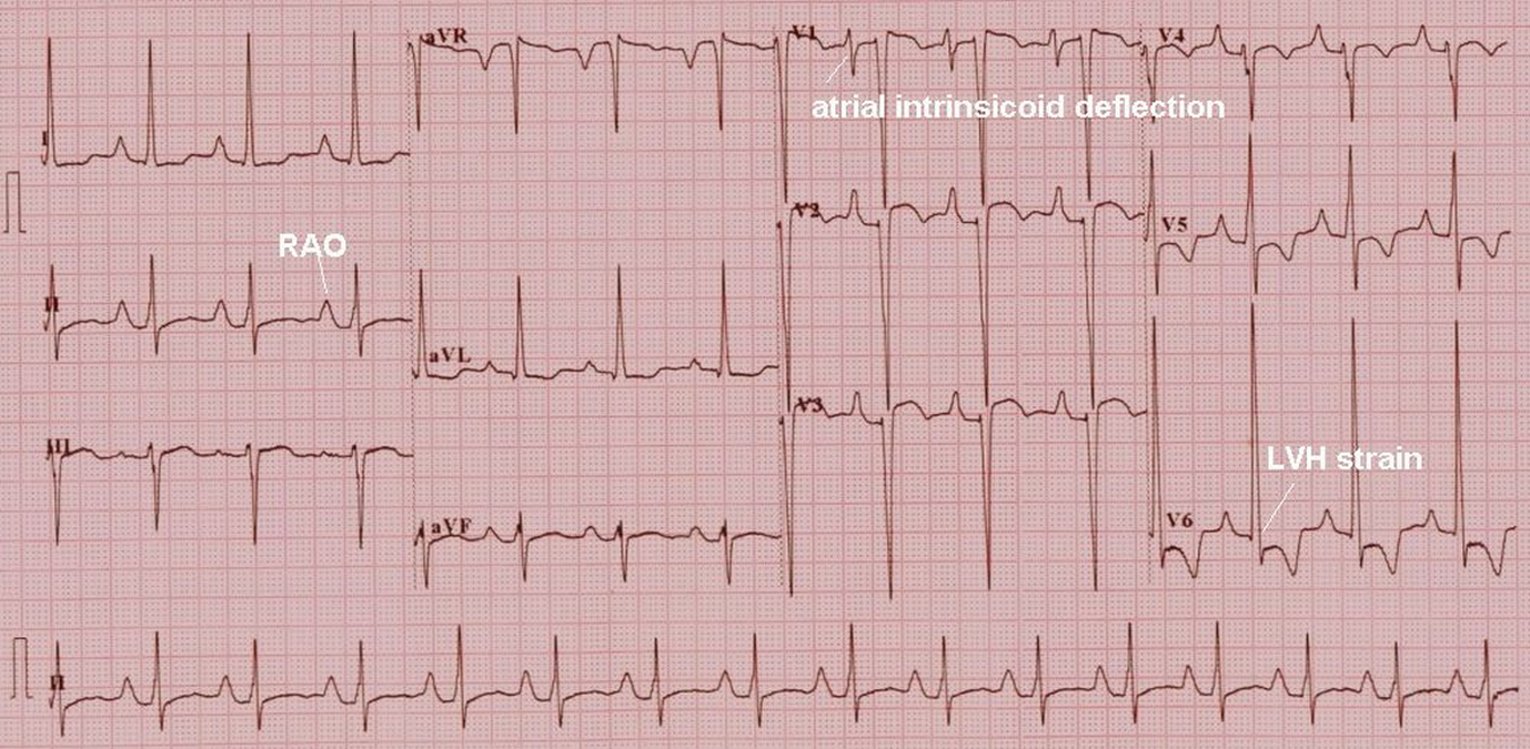
(See annotated image below)
Right atrial overload is manifest as tall P waves in lead II and left ventricular hypertrophy with strain pattern is seen in lateral leads with tall R waves, ST segment depression and T wave inversion. The axis is leftward with predominantly negative QRS in leads III and aVF. The biphasic P wave in V1 with sharp atrial intrinsicoid deflection (the sharp downward deflection from the peak of the P wave to the trough of the P wave) is a pseudo left atrial overload pattern, seen in right atrial overload. In true left atrial overload the atrial intrinsicoid deflection is more slanting so that the negative component of the P wave is almost U shaped rather than the V shape in this case. All these features together in a cyanotic congenital heart disease is characteristic of tricuspid atresia.
Related Posts


About The Author
Johnson Francis
Former Professor of Cardiology, Calicut Govt. Medical Kozhikode, Kerala, India. Editor-in-Chief, BMH Medical Journal