Tissue Doppler Imaging (TDI)
Tissue Doppler Imaging (TDI)
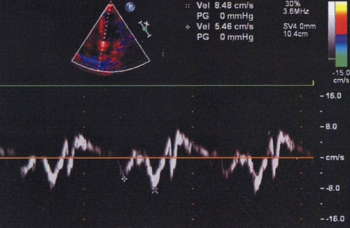
Tissue Doppler Imaging (TDI) measures the velocity of myocardial motion using Doppler principles. While the usual Doppler echocardiography measures the velocity of blood flow using the Doppler signals from the fast moving blood cells, which are of low amplitude, tissue Doppler measures low velocity, high amplitude signals from the myocardial tissue motion. Tissue Doppler is not able to differentiate between passive motion and active motion due to fibre shortening. But the newer technology of strain imaging is able to do so. Colour coded tissue Doppler imaging is sometimes called colour kinesis. Pulsed wave TDI is useful to measure myocardial velocities in the long axis as the movement is parallel to the Doppler beam. The mitral annular TDI has three waves: Sa, systolic myocardial velocity; Ea, early diastolic myocardial velocity and Aa, myocardial velocity during atrial contraction. While imaging from the apical view, systolic velocities are positive and diastolic velocities are negative. Systolic velocity at the lateral mitral annulus correlates with the longitudinal systolic function of the left ventricle. Diastolic velocities depend on ventricular diastolic function. TDI assessment of diastolic function is load independent compared to the conventional measurement using mitral inflow velocities which are highly sensitive to preload.
Related Posts

About The Author
Johnson Francis
Former Professor of Cardiology, Calicut Govt. Medical Kozhikode, Kerala, India. Editor-in-Chief, BMH Medical Journal