Right coronary angiogram
Right coronary angiogram
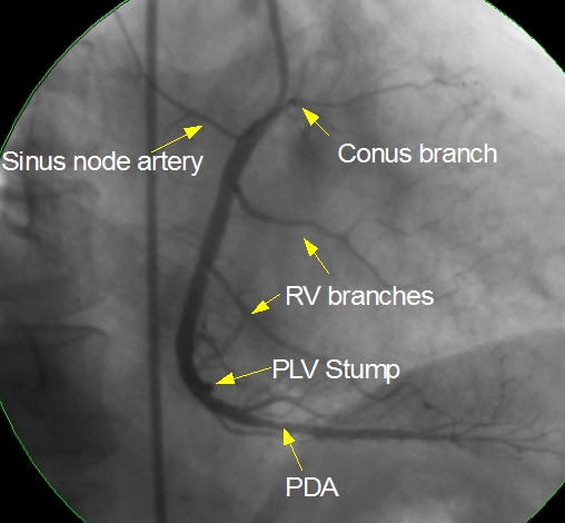
Right coronary angiogram showing conus branch, sinus node artery, right ventricular branches and posterior descending coronary artery. PDA has a mild lesion about 8 mm from its origin. Septal branches can be seen originating from the PDA. Posterior left ventricular branch is occluded soon after its origin and is seen as a stump. Atrioventricular nodal artery usually originates near the bifurcation of the right coronary artery into PDA and PLV. Right coronary angiogram is usually obtained using a Judkins right curve catheter while approaching from the femoral arterial route.
The tip of the JR catheter is seen engaged in the right coronary ostium near the origin of the conus branch. The straight course of the catheter in descending aorta can be seen adjacent the coronary outline. If it had been a transradial approach this would not be visible. Since spine is seen to the right of the image, it is right anterior oblique view. In left anterior oblique view, the spine will be seen to the left of the image. In antero-posterior view, the spine will be in the center.
If the approach is through the radial artery, either the Judkins right catheter or a Tiger catheter can be used. Tiger catheter has the advantage that the same catheter can be used to engage both the right and left coronary ostia. This has the advantage of reducing catheter exchanges in radial artery which is prone for spasm with each catheter exchange.
Related Posts


About The Author
Johnson Francis
Former Professor of Cardiology, Calicut Govt. Medical Kozhikode, Kerala, India. Editor-in-Chief, BMH Medical Journal