Popliteal angiogram
Popliteal angiogram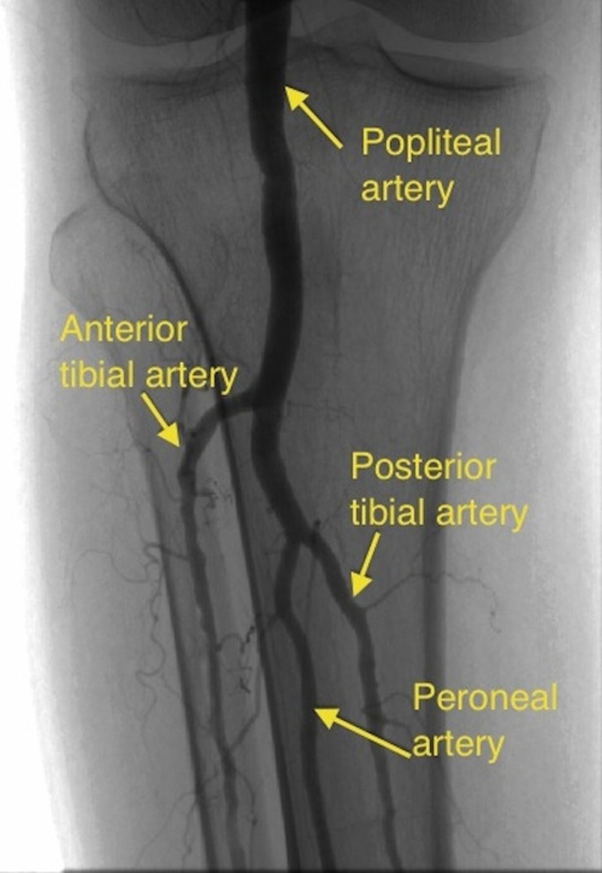
Popliteal angiogram is usually obtained as part of the femoral angiogram by panning the table downwards to cover the popliteal artery as the contrast flows down from the femoral artery. Femoral angiogram can be either obtained by an antegrade puncture of the ipsilateral femoral artery and introducing the sheath downwards in an antegrade fashion or by a catheter introduced through a conventional retrograde puncture of the contralateral femoral artery.
In the latter case, the catheter traverses the contralateral iliac artery to the aortic bifurcation and then down the ipsilateral iliac artery into the femoral artery. This method is used when the ipsilateral puncture is technically difficult. Femoral angiography comes under the broad group of peripheral angiography.
Usually the popliteal artery divides into anterior and posterior tibial branches and the posterior tibial gives off a peroneal branch. Anterior tibial artery becomes the dorsalis pedis artery as it crosses the ankle joint. Posterior tibial artery is palpable posterior to the the medial malleolus. Terminal divisions of posterior tibial artery are the medial and lateral plantar arteries.
Peroneal artery arises from the posterior tibial artery a short distance below its origin. It is also known as the fibular artery. Posterior tibial artery before the origin of peroneal artery is also called the tibioperoneal trunk. Geniculate arteries arise from popliteal artery at multiple levels, providing blood supply to the knee joint and adjacent structures. They also contribute to the collateral blood supply to the lower extremity [1].
Reference
- Zach Bowers Z, Bordoni B. Anatomy, Bony Pelvis and Lower Limb, Popliteal Artery. StatPearls [Internet].
Related Posts

About The Author
Johnson Francis
Former Professor of Cardiology, Calicut Govt. Medical Kozhikode, Kerala, India. Editor-in-Chief, BMH Medical Journal