Pictorial review of CRT implantation
Pictorial review of CRT implantation
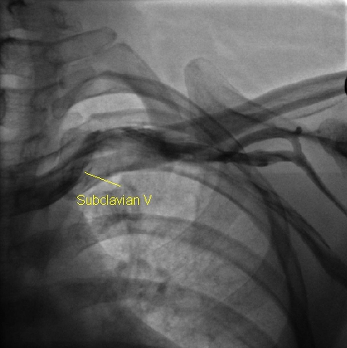 Left subclavian venogram – First step in CRT implantation!
CRT implantation: Left subclavian venogram is usually the first step during the implantation of most cardiac implantable electronic devices (CIED). Iodinated contrast is injected into a left forearm vein and the live fluoroscopy images captured for use as a roadmap during percutaneous subclavian vein puncture for the introduction of intracardiac leads of the device. Patient position on the table is not changed after acquisition of the venogram so that roadmap can be used for fluoroscopy guided subclavian vein puncture. Extrathoracic ‘subclavian’ (axillary) vein puncture is very much facilitated by the venogram roadmap. Intrathoracic puncture is discouraged because of the chance of ‘subclavian crush’ and long term damage to the pacing leads. Intrathoracic subclavian puncture has also a risk of pneumothorax as an occasional complication.
The venogram is also useful to exclude anomalies and occlusion of the venous system prior to puncture. In case the veins are not found to be suitable, procedure is switched to the opposite side.
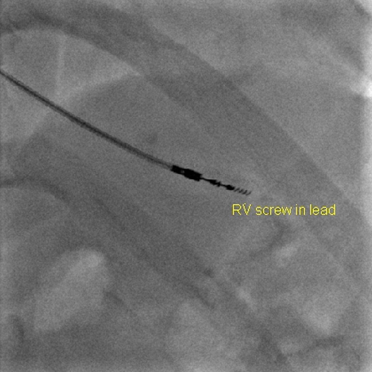 Right ventricular screw in pacing lead
Once the subclavian puncture has been obtained, the right ventricular pacing lead can be introduced and screwed into the desired location. Screw in to the interventricular septum is often preferred to an apical location.
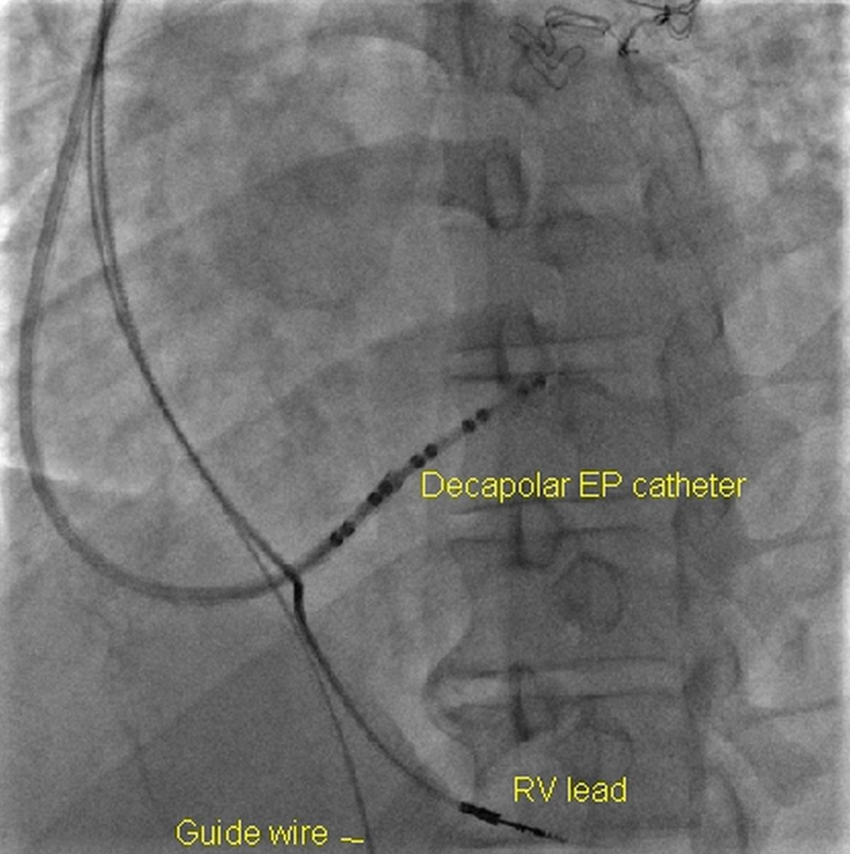 Decapolar EP catheter in the coronary sinus
A steerable decapolar electrophysiology (EP) catheter introduced through a sheath is useful in guiding the sheath to the coronary sinus. Decapolar catheter can be removed once the sheath has been threaded well over it into the coronary sinus and seated well.
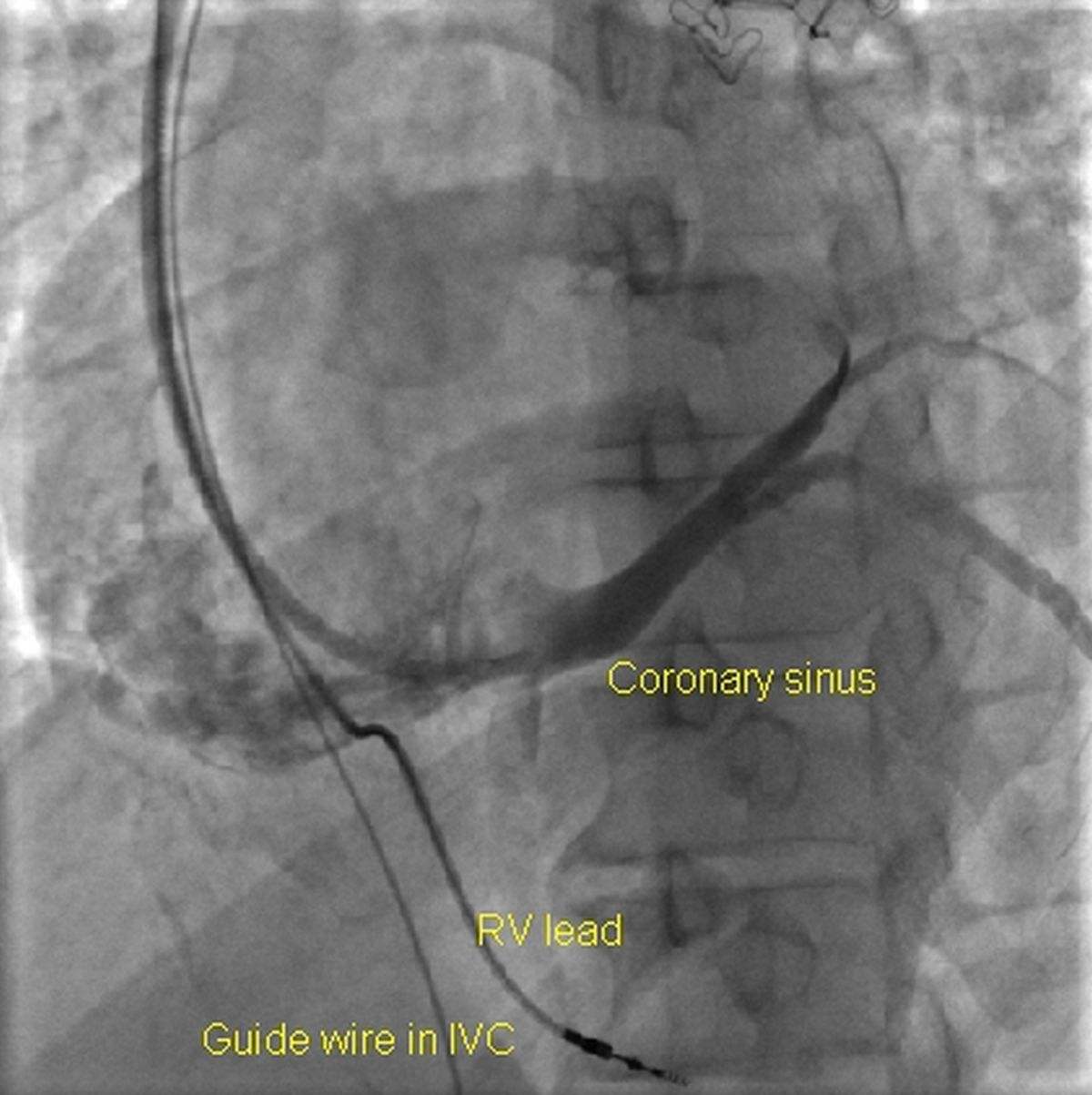 Coronary sinus venogram
Coronary sinus venogram can be obtained by injecting iodinated contrast into the coronary sinus through the sheath. Balloon occlusion of the proximal coronary sinus can improve the visualisation of the coronary venous tree.
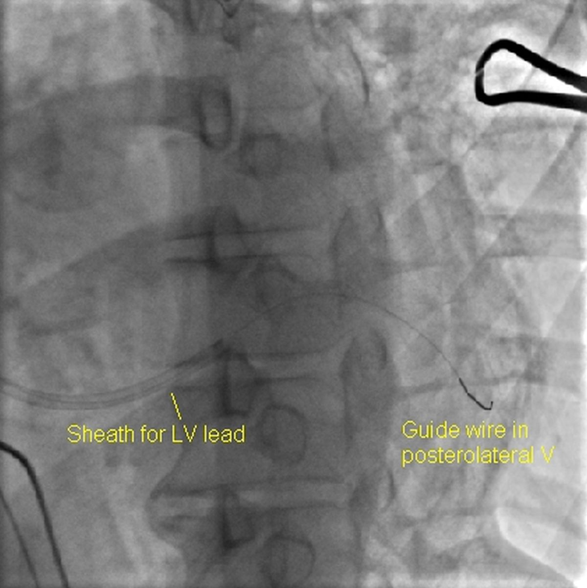 Guidewire in a posterolateral coronary vein
A guidewire can be introduced slowly through the sheath in the coronary sinus and navigated to a good posterolateral vein visualised by the previous coronary sinus venogram.
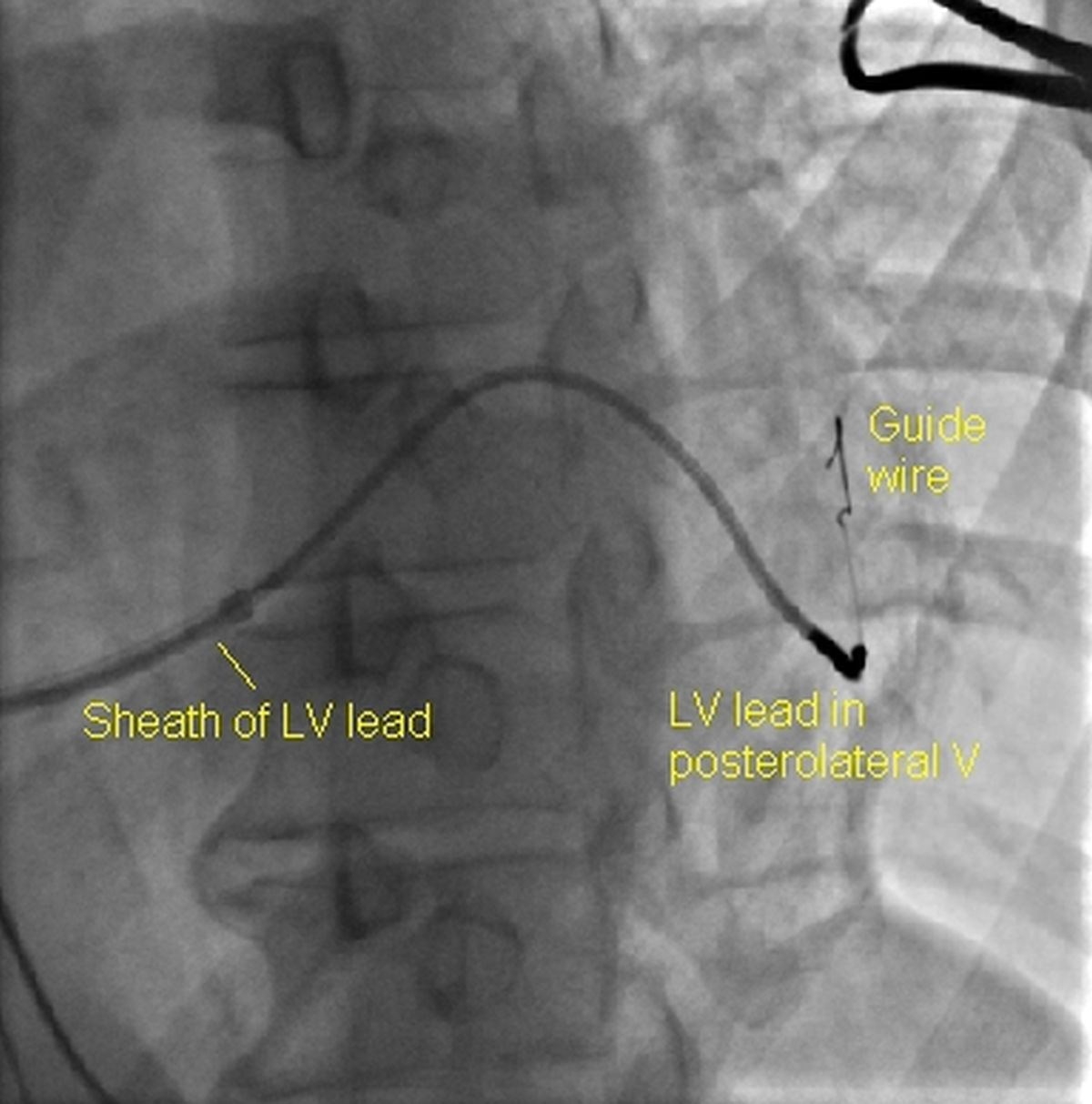 Left ventricular lead in a posterolateral vein
The left ventricular lead of the cardiac resynchronization therapy (CRT) device can be threaded over the guidewire in the posterolateral vein. The peel away sheath can be split using the device provided in the kit once a stable lead position has been obtained and tested.
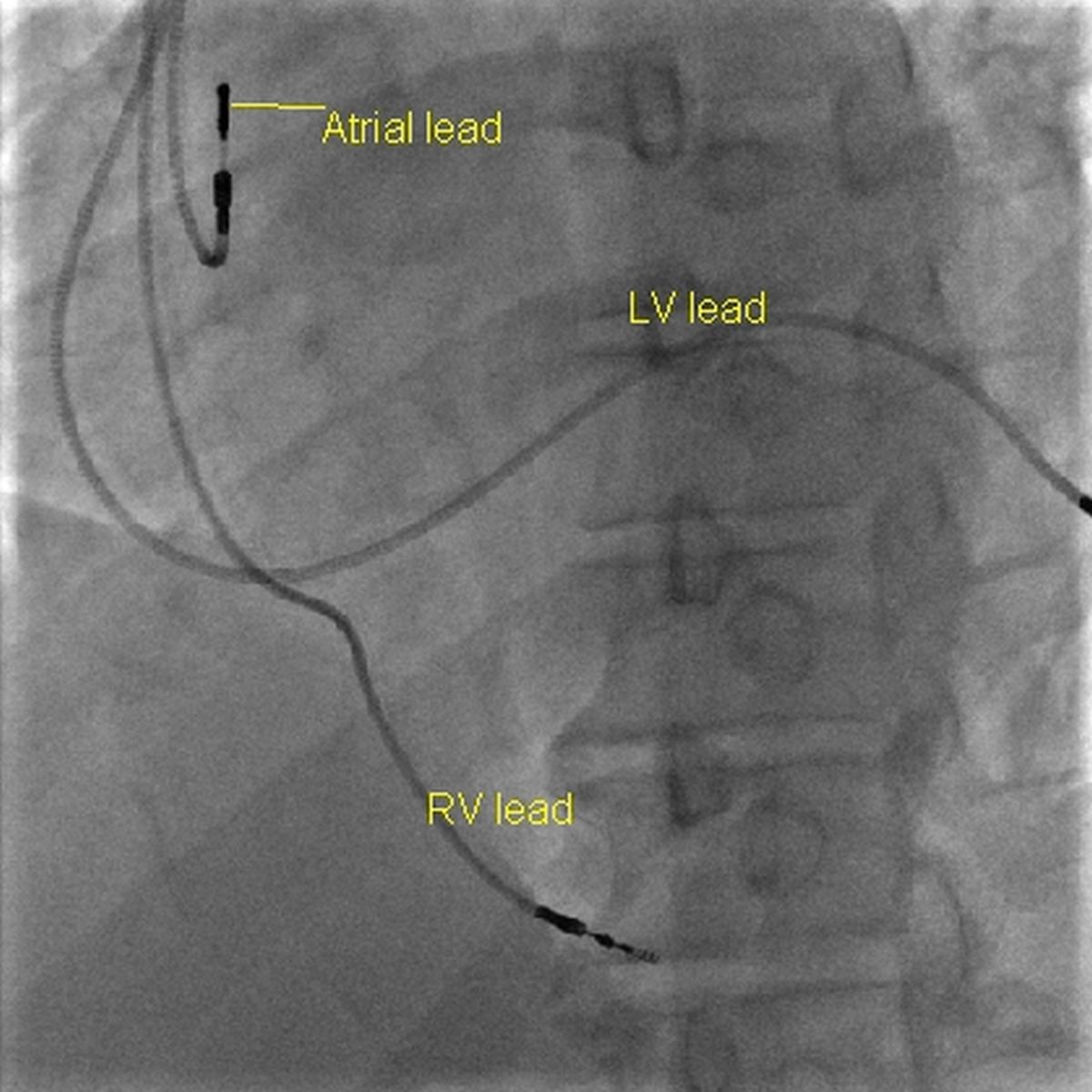 Atrial, right ventricular and left ventricular leads of CRT
Atrial lead can be introduced and positioned in the right atrial appendage. Proximal ends of all the leads are connected to the appropriate ports of the CRT device.
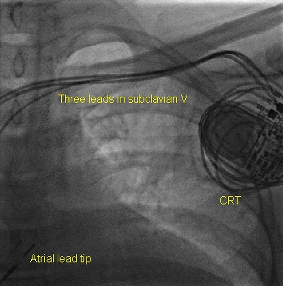 Three leads in left subclavian vein
Three leads are seen in the left subclavian vein. Tip of the atrial lead is seen in the lower right corner. Redundant lead loops and the CRT device are seen in the left upper corner.
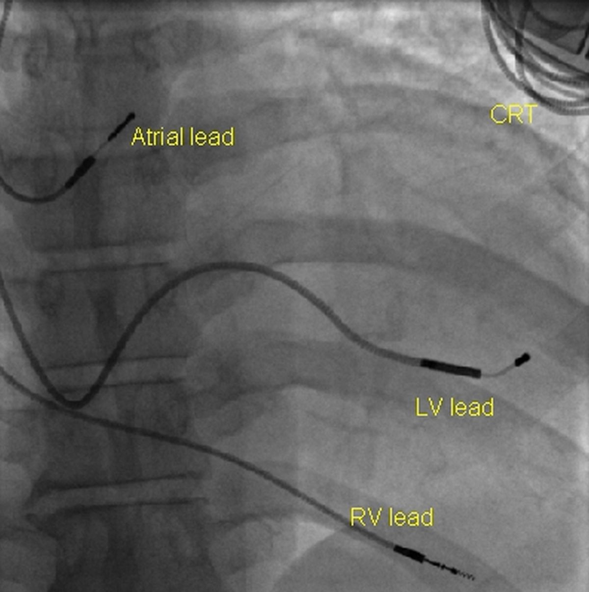 Atrial lead, left ventricular lead and right ventricular leads and part of the CRT
Final picture after implantation of a CRT device showing both ventricular leads, atrial lead and a part of the device in the upper left corner of the image.
Read CRT implantation pearls.
Related Posts


About The Author
Johnson Francis
Former Professor of Cardiology, Calicut Govt. Medical Kozhikode, Kerala, India. Editor-in-Chief, BMH Medical Journal