Latest
ECG / Electrophysiology
ECG showing ventricular ectopics - couplets
Read More
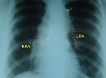
Cardiology X-ray
Dilatation of ascending aorta seen on X-ray chest PA view.
Read More
Cardiology X-ray
Intimal calcification in aortic knuckle noted on Chest X-ray PA view - annotated and unannotated images.
Read More
Angiography and Interventions
Reverse gradient between aorta and left ventricle: can occur rarely in pull back tracings due to peripheral amplification, though theoretically impossible.
Read More
General Cardiology
The various positions of ventricular septal defects in double outlet right ventricle are: Subaortic, subpulmonary, doubly committed, remote or no VSD.
Read More
ECG / Electrophysiology
Clinical types of double outlet right ventricle are TOF like, TGA like, VSD like and Eisenmenger like.
Read More
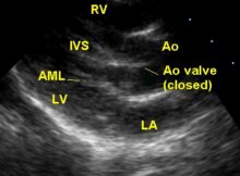
Echocardiogram Library
Echocardiogram with video in parasternal long axis view in Tetralogy of Fallot showing aortic over-ride and ventricular septal defect.
Read More
Echocardiogram Library
Apical five chamber view in Tetralogy of Fallot showing right to left shunt across the ventricular septal defect by color flow mapping.
Read More
General Cardiology
Myocardial glucotoxicity and lipotoxicity in uncontrolled diabetes mellitus may be linked to dilated cardiomyopathy.
Read More
ECG / Electrophysiology
Pacing through lateral cardiac vein showing QS complexes in lead V4-V6 in paced beats indicating an activation proceeding medially from lateral wall of LV.
Read More
Posts navigation