Latest
Echocardiogram Library
Color Doppler echocardiogram showing right to left shunt across a subaortic ventricular septal defect
Read More
Echocardiogram Library
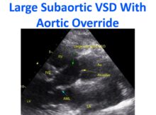
Large subaortic VSD with aortic override - echocardiogram, seen from the parasternal long axis view
Read More
Echocardiogram Library
Trivial aortic regurgitation - echocardiogram seen on colour Doppler echocardiogram in the left image (AR)
Read More
ECG / Electrophysiology
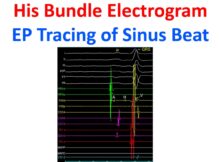
EP tracing of sinus beat - His bundle electrogram
Read More
Cardiac CT scan
Cob web sign in aortic dissection are slender linear areas of low attenuation seen in false lumen in contrast enhanced computerized tomographic (CT) scans
Read More
ECG / Electrophysiology
Channels of conduction within scars of myocardial infarction, if connected to normal myocardium, can be the source of ventricular arrhythmias.
Read More
Angiography and Interventions
Non dominant left circumflex coronary artery seen on left coronary angiogram in right anterior oblique (RAO) caudal view.
Read More
Angiography and Interventions
Optimal time for elective non cardiac surgery after coronary stenting is between 46 - 180 days for bare metal stents, after 180 days for drug eluting stents
Read More
Angiography and Interventions
Left anterior oblique (LAO) caudal view of left coronary angiogram resembles a spider and hence the term spider view.
Read More
Echocardiography
Yu index of longitudinal tissue Doppler dyssynchrony is the standard deviation of time to peak systolic velocities from the onset of the QRS complex.
Read More
Posts navigation