Mitral E/E’ ratio on echocardiogram
Mitral E/E’ ratio on echocardiogram
Abstract: Mitral E/E’ ratio on echocardiogram is an important indicator of left ventricular diastolic function. It can be used in the presence of atrial fibrillation when E/A ratio is not available.
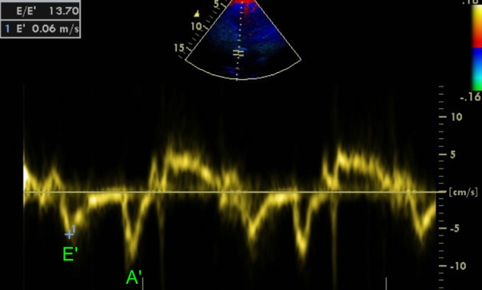
Tissue Doppler echocardiography showing measurement of mitral E/E’ ratio for assessment of diastolic dysfunction. Initially mitral E wave is measured and stored as given in the image below. After that colour Tissue Doppler (tissue velocity imaging or TVI) mode is switched on to assess tissue Doppler. The cursor is placed over the medial mitral annulus and tissue Doppler tracing obtained. E’ velocity is measured and stored. The software usually gives the E/E’ ratio. In this case the ratio is given as 13.7, suggesting diastolic dysfunction. It may be noted that the Nyquist limit for the colour tissue Doppler is 0.16 meters per second in this image while it is 0.65 meters per second in the image below for colour Doppler imaging (colour flow mapping). This is because the blood flow velocities are much higher than the velocity of tissue movement with systole and diastole.
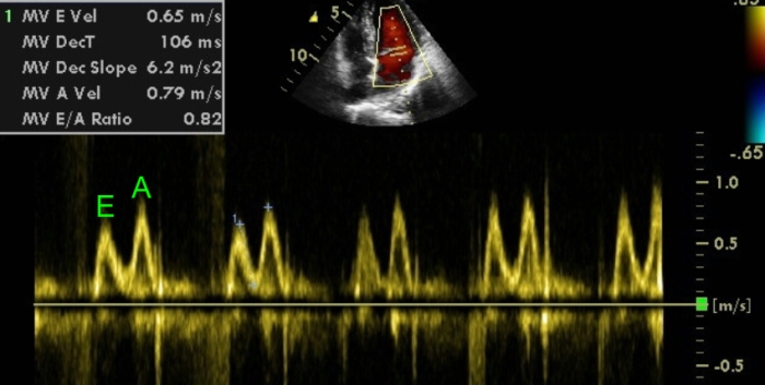
Conventional Doppler assessment of transmitral flow showing E/E reversal in left ventricular diastolic dysfunction. MV E Vel: early mitral inflow velocity; MV DecT: deceleration time of the early mitral inflow velocity; MV Dec Slope: deceleration slope of mitral inflow velocity; MV A Vel: mitral inflow velocity during atrial systole; MV E/A Ratio: ratio between early mitral inflow velocity and atrial systolic mitral inflow velocity.
Limitations of mitral inflow velocity in left ventricular diastolic function assessment
Even though the most widely used measure of left ventricular diastolic dysfunction is documentation of E/A reversal in mitral inflow velocity by Doppler echocardiography, it has several limitations. Mitral inflow is dependent on the rate of ventricular relaxation, left atrial pressure, relative compliance of atrial and ventricular chambers as well as the ventricular suction effect [1].
Tissue Doppler assessment of myocardial velocity for assessment of left ventricular diastolic function
Tissue Doppler assessment of myocardial velocity is useful in assessment of left ventricular diastolic function. Ratio of the Doppler derived E to tissue Doppler derived E’ (E/E’) has been shown to be a good predictor of mean left ventricular diastolic pressure. Eighty five percent of those with an E/E’ ratio of less than 8 had normal mean left ventricular diastolic pressures. All of those who had E/E’ more than 15 had elevated mean left ventricular diastolic pressures.
E/E’ ratio has the advantage that it can be used even in the presence of atrial fibrillation where E/A ratio is not available. To enhance the accuracy, determination of E/E’ ratio in a single beat using dual Doppler echocardiography has also been reported [2].
Reference
- Mottram PM, Marwick TH. Assessment of diastolic function: what the general cardiologist needs to know. Heart. 2005 May;91(5):681-95.
- Kusunose K, Yamada H, Nishio S, Tomita N, Niki T, Yamaguchi K, Koshiba K, Yagi S, Taketani Y, Iwase T, Soeki T, Wakatsuki T, Akaike M, Sata M. Clinical utility of single-beat E/e’ obtained by simultaneous recording of flow and tissue Doppler velocities in atrial fibrillation with preserved systolic function. JACC Cardiovasc Imaging. 2009 Oct;2(10):1147-56.
Related Posts


About The Author
Johnson Francis
Former Professor of Cardiology, Calicut Govt. Medical Kozhikode, Kerala, India. Editor-in-Chief, BMH Medical Journal