Mesocardia with L-TGA
Mesocardia with L-TGA
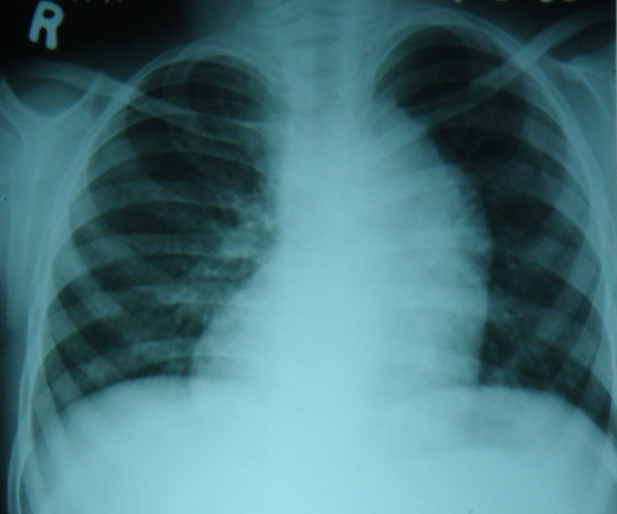
The cardiac shadow is seen in the midline, with almost equal spread to either side, suggesting mesocardia. Mesocardia means a centrally located heart [1]. In levocardia the heart is predominantly in the left hemithorax and the apex points to the left. In dextrocardia, the heart is predominantly in the right hemithorax and the apex points to the right.
Normally the aorta is dextro-posed and the ascending aorta is seen along the right cardiac border. In levo-posed (L-posed) aorta, the ascending aorta is on the left border, as in this case. In dextro-transposition of great arteries (D-TGA), aorta is dextro-posed and in levo transposition of great arteries, aorta is levo-posed.
D-transposition is the usual transposition of great arteries, which is a cyanotic congenital heart disease with increased pulmonary blood flow (unless there is associated severe pulmonary stenosis – left ventricular outflow tract obstruction). L-transposition is usually a corrected transposition of great arteries with atrioventricular and ventriculoarterial discordance so that right atrial blood reaches the pulmonary artery and left atrial blood reaches the aorta. Hence it is a non cyanotic condition and may be missed unless there are other associated anomalies like ventricular septal defect, pulmonary stenosis or congenital complete heart block, which are the common associations of L-TGA.
Important differential diagnosis for a shadow along the left upper cardiac border are:
- L – posed aorta (levo posed aorta)
- Dilated main pulmonary artery
- Aneurysm of arch or upper descending thoracic aorta
- Vertical vein in total anomalous or hemi-anomalous pulmonary venous connection
- Aneurysm of the ductus arteriosus
- Partial absence of left pericardium causing bulge of cardiac structures to the left
- Left atrial appendage
- Submitral aneurysm (a little lower on the silhouette)
In a study assessing the laterality relationship in transposition of great arteries, 11 cases of mesocardia were seen [2]. Six of them had usual abdominal organ arrangement while 4 had mirror-image arrangement. One had right isomerism.
25% of cases of corrected transposition of great arteries have either dextrocardia or mesocardia [3]. Echocardiography is difficult in mesocardia because majority of the heart is beneath the sternum. Precordial echo windows have limited penetration. Subcostal and suprasternal windows are useful in infants while transesophageal echo is useful in adults.
References
- Van Praagh R. Terminology of congenital heart disease. Glossary and commentary. Circulation. 1977 Aug;56(2):139-43.
- Al-Zahrani RS, Alharbi SH, Tuwaijri RMA, Alzomaili BT, Althubaiti A, Yelbuz TM. Transposition of the great arteries: A laterality defect in the group of heterotaxy syndromes or an outflow tract malformation? Ann Pediatr Cardiol. 2018 Sep-Dec;11(3):237-249.
- Chaudhuri M, Tomar M. Echocardiographic Approach to Congenitally Corrected Transposition. J Indian Acad Echocardiogr Cardiovasc Imaging 2020;4:312-24.
Related Posts

About The Author
Johnson Francis
Former Professor of Cardiology, Calicut Govt. Medical Kozhikode, Kerala, India. Editor-in-Chief, BMH Medical Journal