Low atrial rhythm and IVCD
Low atrial rhythm and IVCD
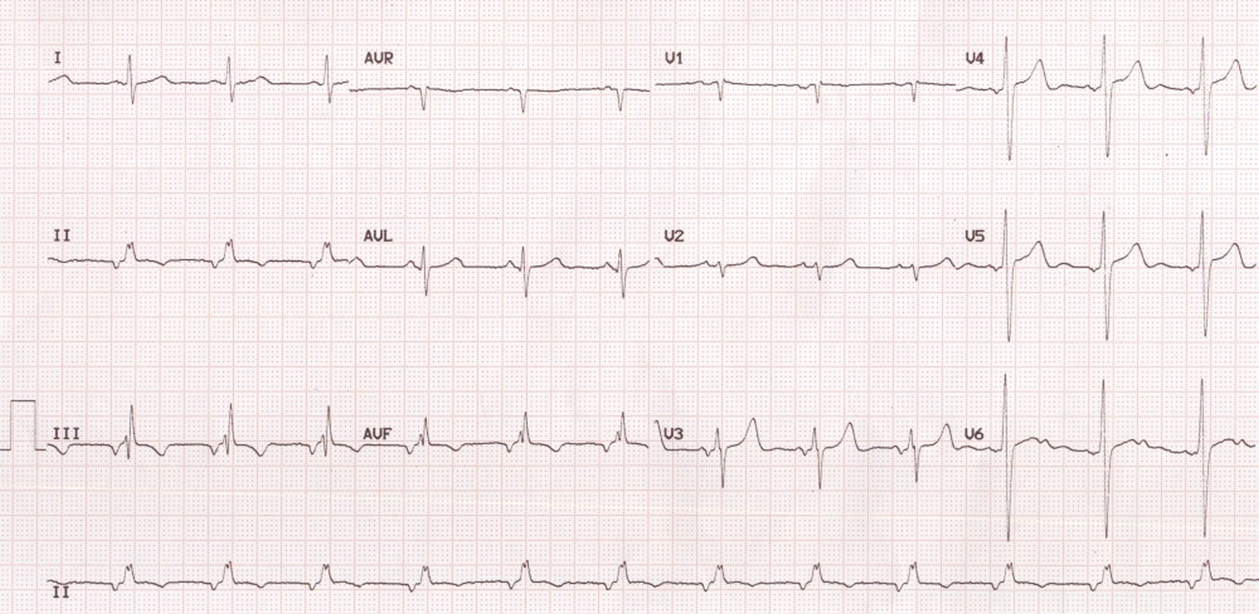
ECG showing negative P waves in inferior leads – II, III and aVF. A superior P wave axis means that the atrial activation is proceeding from below upwards. This occurs when the focus is in the low atrium (low atrial rhythm or coronary sinus rhythm). In addition the ECG also shows an intraventricular conduction defect (IVCD) in the form of notched R wave in lead II and aVF and an rSR’ pattern in lead III. Sometimes this pattern occurs in atrial septal defect and it is known as crochetage sign. The QRS width seems to be a little more than that in usual crochetage sign. (Low atrial rhythm can occur in sinus venosus ASD as the region of the sinus node is defective and an ectopic atrial focus takes over.
Related Posts


About The Author
Johnson Francis
Former Professor of Cardiology, Calicut Govt. Medical Kozhikode, Kerala, India. Editor-in-Chief, BMH Medical Journal