LV aneurysm – left ventriculogram
LV aneurysm – left ventriculogram
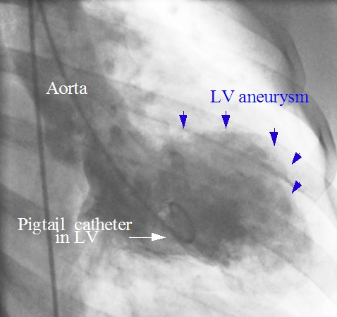
This still picture from a left ventricular angiogram shows a bulge in the anterolateral region of the left ventricle in systole suggestive of an aneurysm. A is composed of all three layers of the heart, namely endocardium, myocardium and pericardium. It has a wide neck and is very unlikely to rupture. In contrast, a pseudoaneurysm is a ventricular rupture sealed off by the pericardium. It has a narrow neck and has a high risk of secondary rupture. A true aneurysm can cause arrhythmias due to the viable tissue in the border zone with varying refractory periods which initiate re-entrant arrhythmias. Refractory heart failure may occur due to wasted systole – i.e., a portion of the ventricular blood moves in and out of the aneurysm without being pumped out into the aorta.
Huge left ventricular aneurysm with thrombus and calcification has been reported by de Agustin JA et al [1]. The patient presented with chronic heart failure after a previous myocardial infarction. Echocardiogram and left ventriculogram confirmed a giant calcified anteroapical left ventricular aneurysm with very severe left ventricular systolic dysfunction. The patient received an implantable cardioverter defibrillator for primary prevention. Thromboembolism is another potential problem with left ventricular aneurysm containing thrombus.
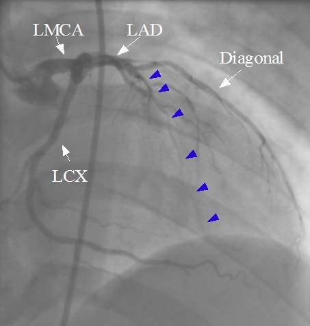
The region of the aneurysm is usually the territory of a poorly collateralized total or near total coronary occlusion. In this case it is near total occlusion of the left anterior descending (LAD) coronary artery beyond a major diagonal with faint antegrade flow (blue arrows).
Surgical repair of the aneurysm is done by left ventricular reconstruction [2]. This is done after complete revascularization by coronary artery bypass grafting. Left internal mammary artery and if necessary, saphenous vein grafts are used for complete revascularization. Mitral valve repair is also done along with if deemed necessary. Surgical left ventricular reconstruction is an optional procedure to improve the left ventricular volume and shape and possibly long term improvement in left ventricular function. The scar tissue is excluded in this open procedure. Though several studies have reported that surgical left ventricular reconstruction is safe and has favorable five year outcomes, the need is still being debated.
References
- de Agustin JA, de Diego JJ, Marcos-Alberca P, Rodrigo JL, Almeria C, Mahia P, Luaces M, Garcia-Fernandez MA, Macaya C, de Isla LP. Giant and thrombosed left ventricular aneurysm. World J Cardiol. 2015 Jul 26;7(7):431-3.
- Castelvecchio S, Menicanti L. Left ventricular reconstruction: update to left ventricular aneurysm/reshaping techniques. Multimed Man Cardiothorac Surg. 2013;2013:mmt002. doi: 10.1093/mmcts/mmt002.
Related Posts


About The Author
Johnson Francis
Former Professor of Cardiology, Calicut Govt. Medical Kozhikode, Kerala, India. Editor-in-Chief, BMH Medical Journal