Femoral artery calcification
Femoral artery calcification
Interesting X-ray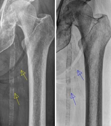
Femoral artery calcification – negative and positive images
Incidentally detected femoral artery calcification on X-ray taken for some other purpose. Both positive and negative images are shown side by side. Positive images were obtained by digital processing of the negative image. Extensive calcification is seen extending from the common femoral artery into superficial femoral artery and profunda femoris artery.
Arterial calcifications are more likely to be seen in the elderly and in those with altered calcium metabolism as in chronic kidney disease and hyperparathyroidism. Secondary hyperparathyroidism may occur in chronic kidney disease due to elevated levels of parathormone which was even considered as a uremic toxin.
It can be seen that upper part of the common femoral artery passes over the femoral head, the site where it is commonly punctured by Seldinger technique for angiography, insertion of intra arterial balloon pump and sometimes even for arterial blood gas sampling. It may be noted that femoral puncture for ABG is used only if radial access is not feasible, to avoid unnecessary trauma to femoral artery by frequent use for ABG.
The close relation of the femoral artery in the anteroposterior view to the femoral head is utilized for fluoroscopy guided puncture in case of obese individuals when pulsations of the femoral artery are not easily felt for guiding blind percutaneous puncture. Caution is needed in using fluoroscopic guidance often as it entails extra radiation to the operators hand as well.
Related Posts

About The Author
Johnson Francis
Former Professor of Cardiology, Calicut Govt. Medical Kozhikode, Kerala, India. Editor-in-Chief, BMH Medical Journal