EP tracing of sinus beat – His bundle electrogram
EP tracing of sinus beat – His bundle electrogram


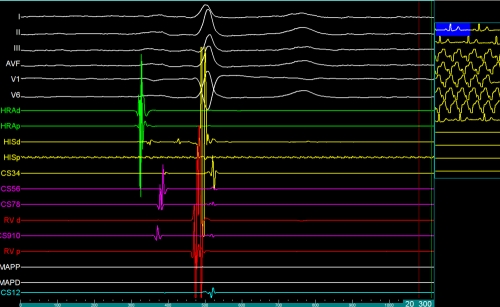
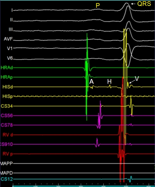
EP tracing of a sinus beat along with multiple surface ECG recordings. Surface ECG recordings are from leads I, II, III, AVF, V1 and V6. The P wave and QRS complex have been marked. The sweep speed is much higher than that for conventional ECG recordings. Calibration in milliseconds can be seen in the thin green panel at the bottom (0, 100, 200, 300, 400, 500, 600). A: atrial complex in the His bundle electrode; H: classical triphasic His bundle potential; V: ventricular potential recorded from the His bundle electrode. HRAd: recording from the distal pair of high right atrial electrodes;
HRAp: recording from the proximal pair of high right atrial electrodes; CS12: recording from the distal most pair of coronary sinus electrodes; CS34, CS56, CS78 and CS910 are the serially proximal recordings from the the coronary sinus electrodes in the decapolar catheter. CS12 usually represents left atrial recording from distal coronary sinus. RVp: proximal pair of right ventricular electrodes; RVd: distal pair of right ventricular electrodes;
MAPD: distal pair of mapping electrodes, the ablation catheter used for delivering radiofrequency energy; MAPP: proximal pair of mapping electrodes. Please note that atrial recordings in the high right atrial tracing occurs earlier than those in the coronary sinus. Proximal to distal propagation can be noted in the coronary sinus electrodes, indicating normal activation pattern which spreads from the high right atrium down and left.


