Echocardiographic profile of prosthetic mitral valve
Echocardiographic profile of prosthetic mitral valve
This echocardiographic profile of prosthetic mitral valve with series of images can be considered as a useful baseline information while imaging prosthetic valves which is often challenging due to the dense acoustic shadowing by the prosthetic material.
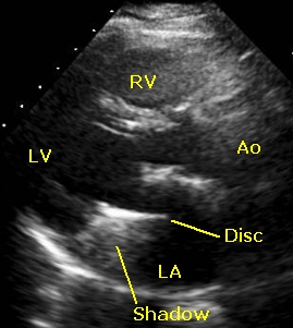
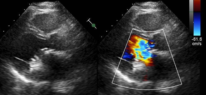
Usually we start imaging from the parasternal long axis view (PLAX). In this view we can see the outflow region of the right ventricle (RV), basal part of the left ventricle (LV) with the interventricular septum in between (unmarked). Left ventricular outflow tract (unmarked) above the mitral valve can be seen leading to the aorta (Ao). Left atrium (LA) is seen posterior to the aorta. The prosthetic mitral valve disc (disc) is seen in a horizontal position here with significant echo density of the metallic component. Acoustic shadowing (shadow) is seen posterior to the dense disc shadow, which extends up to the posterior wall of the left atrium, proximal to the mitral annulus. Because of the acoustic shadowing, it will be difficult to recognize any structure in that region like a vegetation or thrombus.
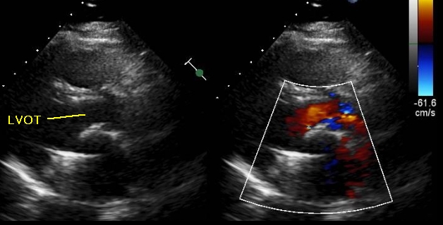
This split screen image shows 2-dimensional (2D) image on the left side and colour Doppler in the right panel. With the disc in horizontal position, there is no colour flow across the mitral valve. At the same time red coloured flow is seen anterior to the mitral valve in the left ventricular outflow tract (LVOT) and into the aorta.
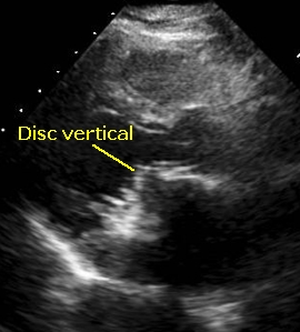
In this image from the PLAX view tilting mitral prosthetic disc is seen in the vertical position, with a slight gap from anterior part of the annulus. It is in a partially open position.
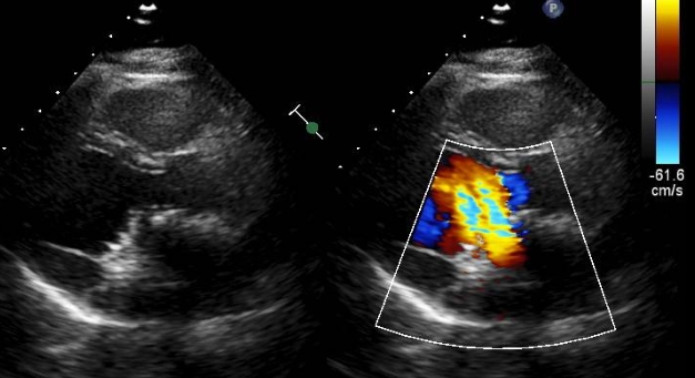
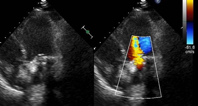
Split image with disc vertical and a a good gap seen between the disc and anterior mitral annulus region in the left panel. Right panel shows excellent colour flow across the mitral prosthetic valve with mild turbulence, which is usual across prosthetic valves. Flow is seen from left atrium into the left ventricle in an antero apical direction.
Another sequence from the same view in which the 2-D image shows better separation of the disc from the anterior annulus and a good flow across the mitral valve in the right panel wih colour flow mapping (colour Doppler).
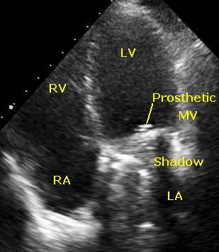
Apical four chamber view showing prosthetic mitral valve (Prosthetic MV) with acoustic shadowing proximal to it in the left atrium (LA). Left ventricle (LV), right ventricle (RV) and right atrium (RA) are also seen. Interventricular septum is seen between the two ventricles (unmarked) and the interatrial septum between the two atria (unmarked).
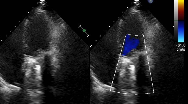
Apical 4C view with disc in the closed position (left panel) and Colour Doppler (right panel) showing no flow across. Acoustic shadowing proximal to the prosthetic mitral valve in the left atrium is seen quite prominently.
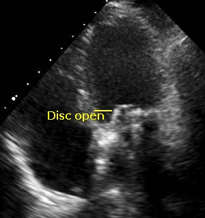
Apical 4C view with the tilting disc of the prosthetic mitral valve in the partially open position. Short area of echo lucency to the right of the disc is seen.
Split screen image from the apical 4C view with left panel showing the disc in open position and right panel showing the flow across the prosthesis on color Doppler.
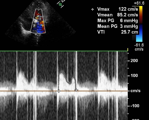
Continuous wave (CW) Doppler interrogation across the prosthetic mitral valve showing the transmitral gradient (sketched out), with peak gradient of 6 mm Hg and mean gradient 3 mm Hg which are within normal limits. Please note the dense vertical shadows on either side of mitral Doppler tracing which is the artefact due to the prosthetic valve opening and closing.
Related Posts


About The Author
Johnson Francis
Former Professor of Cardiology, Calicut Govt. Medical Kozhikode, Kerala, India. Editor-in-Chief, BMH Medical Journal