Echocardiogram in ventricular septal defect (VSD)
Echocardiogram in ventricular septal defect
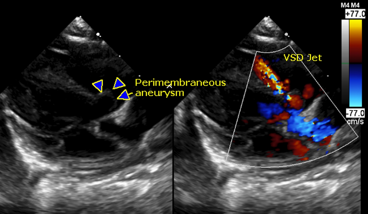
Echocardiogram in ventricular septal defect: Parasternal long axis view shows the subaortic perimembranous ventricular septal aneurysm (marked by arrows). There is a small ventricular septal defect at the apex of the aneurysm which is not very clear in the 2D (two dimensional) image. Right panel shows the mosaic (red multicoloured) VSD jet from the apex of the aneurysm into the right ventricle. Other structures seen in the image (not marked are the aorta (with blue colour flow mapping in right panel), left ventricle (to the left and below the VSD jet) and the left atrium (below and to the left of aortic blue jet). Mitral valve in the closed position between the left atrium and left ventricle (the frame in which VSD jet is visible will be a systolic frame).
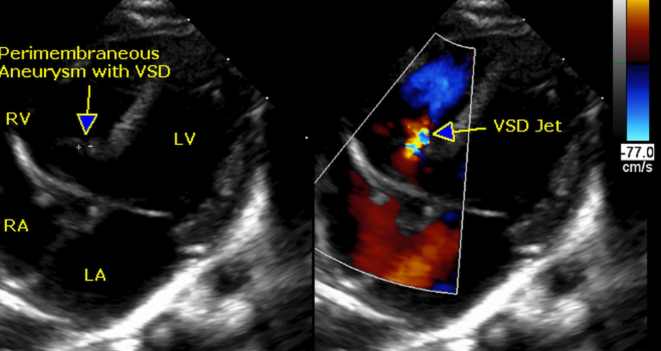
Echocardiogram in apical four chamber (slightly tilted) showing all four cardiac chambers (RA: right atrium; RV: right ventricle; LA: left atrium; LV: left ventricle) and the perimembranous ventricular septal aneurysm with defect (arrow) in the left panel. Right panel shows the reddish mosaic (multi colored) jet through the ventricular septal defect.
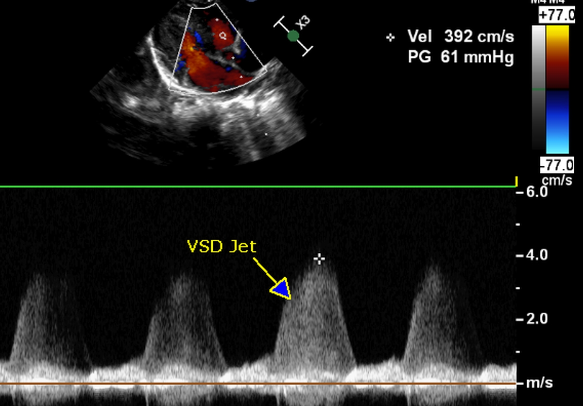
Continuous wave (CW) Doppler interrogation of the ventricular septal defect showed a pansystolic jet with a peak gradient of 61 mm Hg. This will be the difference between the systolic pressures of left and right ventricle. In a large VSD, when the pressures in the two ventricles are more or less equal, the velocity of the VSD jet will be small and hence the gradient will be low. When the pressure gradient across the VSD is high, it usually means that the VSD is restrictive. Here it is a restrictive VSD. Sometimes the right coronary cusp prolapse can cause aortic regurgitation even in a restrictive VSD [1].
Reference
- Abqari S, Rabbani MU, Meshram HS, Gupta A. RCC prolapse causing Aortic regurgitation in a restrictive VSD. Images Paediatr Cardiol. 2015 Jan-Mar;17(1):4-6.
About The Author
Johnson Francis
Former Professor of Cardiology, Calicut Govt. Medical Kozhikode, Kerala, India. Editor-in-Chief, BMH Medical Journal