Ebstein’s Anomaly of Tricuspid Valve
Ebstein’s Anomaly of Tricuspid Valve
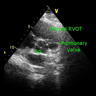
Parasternal short axis view in Ebstein’s anomaly, showing the dilated right ventricular outflow tract (RVOT). Ao: Aorta.
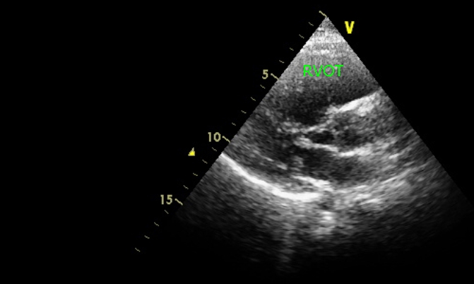
Parasternal long axis view in Ebstein’s anomaly showing the dilated right ventricular outflow tract (RVOT).
Dilated RVOT can be appreciated clinically as a wavy pulsation in the parasternal region which is better seen than felt. There is no sustained heave as with the left parasternal heave of right ventricular hypertrophy. Dilated RVOT is a feature seen in right ventricular endomyocardial fibrosis (RVEMF) as well. In that case there is no displacement of tricuspid valve. Instead there is fibrosis of right ventricular apex.
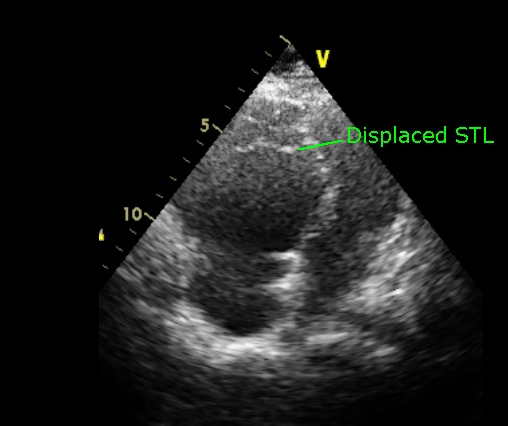
Apical four chamber view in Ebstein’s Anomaly showing the grossly displaced septal tricuspid leaflet (STL). Normal distance between the septal attachments of septal tricuspid leaflet and anterior mitral leaflet is only about 5 millimeters. The portion of right ventricle proximal to the displaced septal tricuspid leaflet is considered to be ‘atrialized’, with a pressure tracing showing atrial pattern and an electrode catheter showing ventricular activity (Hernandez sign) [1,2].
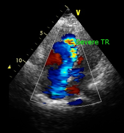
Apical four chamber view in Ebstein’s anomaly showing severe tricuspid regurgitation (TR).
References
- Hernandez FA, Rochkind R, Cooper HR. The intracavitary electrocardiogram in the diagnosis of Ebstein’s anomaly. Am J Cardiol. 1958 Feb;1(2):181-90.
- Watson H. Electrode catheters and the diagnosis of Ebstein’s anomaly of the tricuspid valve. Br Heart J. 1966 Mar;28(2):161-71.
Related Posts

About The Author
Johnson Francis
Former Professor of Cardiology, Calicut Govt. Medical Kozhikode, Kerala, India. Editor-in-Chief, BMH Medical Journal