Fine atrial fibrillation
Fine atrial fibrillation
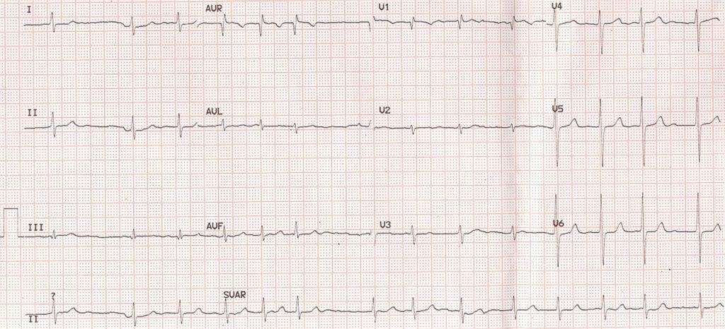
Fine atrial fibrillation is AF in which fibrillary waves are fine (less than 0.5 mm or hardly visible sometimes).
Coarse AF: Fibrillary waves usually more than 1 mm, indicating atrial enlargement or hypertrophy. In atrial fibrillation there is no effective contraction of the atria.
The fibrillary waves are usually of low amplitude and many of them do not get conducted to the ventricles due to refractoriness of the AV node. Frequency of the fibrillary waves may be anything from 400-600 /minute. But the ventricular rate is usually around 120 per minute and irregular. If the AV node is diseased or blocked by drugs, the ventricular rate may be slow.
The fibrillary waves may be coarse if the atria are enlarged as in mitral stenosis. Amplitude of fibrillary waves more than 1 mm is taken as evidence of atrial enlargement. If the ventricular rhythm is regular in atrial fibrillation, it signifies complete AV block and the ventricle being controlled by a junctional or ventricular focus. Usually the QRS complex is narrow in atrial fibrillation.
If the QRS is wide, it could be due to preexisting bundle branch block, aberrancy or pre-excitation. In pre-excitation with atrial fibrillation, the ventricular rate may go as high as 300 per minute and degenerate into ventricular fibrillation. This is because accessory pathways may conduct at very high rate as the refractory period decreases with increasing rate, unlike in AV node.
Important causes of atrial fibrillation include mitral valve disease, coronary artery disease, cardiomyopathy (dilated, hypertrophic and restrictive) and hyperthyroidism. Atrial fibrillation can also occur following binge drinking, which is known as holiday heart syndrome.
An important differential diagnosis of atrial fibrillation with a slow ventricular rate is sinus arrhythmia. In sinus arrhythmia, though the rhythm is irregular, each QRS will be preceded by a P wave and the rate change is related to the respiratory cycle. The cyclical pattern is evident on the ECG monitor which shows a faster rate in inspiration and slower rate in expiration.
Related Posts

About The Author
Johnson Francis
Former Professor of Cardiology, Calicut Govt. Medical Kozhikode, Kerala, India. Editor-in-Chief, BMH Medical Journal