Arterial tracing in supraventricular ectopy
Arterial tracing in supraventricular ectopy
Arterial tracing in supraventricular ectopy
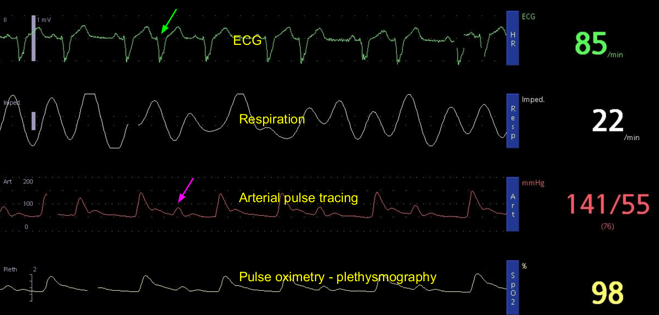
This arterial tracing in supraventricular ectopic along with ECG, respiration and pulse oximetry is useful in learning certain hemodynamic principles. Supraventricular ectopic is marked with green arrow in the top tracing which shows the ECG. Though the QRS complex is wide, it is a supraventricular ectopic beat because the QRS complex resembles the other QRS complexes. Ideally simultaneous multi lead ECG monitoring is needed to confirm that it is really a supraventricular ectopic.
In a single lead, ventricular ectopy and supraventricular ectopy with pre-existing intraventricular conduction defect as in this case, could have similar morphology. But if multiple leads show same morphology for sinus beats and ectopic beats, then it is most likely a supraventricular ectopic beat. Close scrutiny of the descending limb of the preceding T wave shows a small hump which could be a direct indication of the supraventricular ectopic P wave.
Since the arterial tracing is from the radial artery, a significant delay can be noted from the onset of the QRS complex to the onset of the upstroke of the arterial wave. This delay will include both the electromechanical delay of the ventricle and the time taken for the pulse wave to travel to the radial artery cannula.
The lower amplitude of the arterial tracing of a supraventricular ectopic beat (pink arrow) is quite evident. This is because of a shortened preceding diastole. Shortened preceding diastole because of prematurity of the beat leads to poor ventricular filling and hence a lower output. Details about the other tracings can be read from a previous post on arterial tracing in sinus rhythm.
Related Posts


About The Author
Johnson Francis
Former Professor of Cardiology, Calicut Govt. Medical Kozhikode, Kerala, India. Editor-in-Chief, BMH Medical Journal