12 lead ECG – Normal ECG
12 lead ECG – Normal ECG
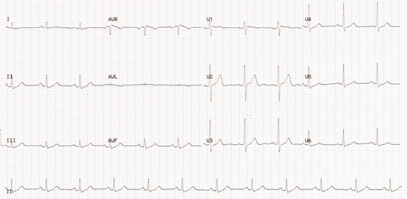
12 lead ECG consists of three standard limb leads (Leads I, II and III), three augmented limb leads (aVR, aVL, and aVF) and six chest leads (V1 to V6). All the twelve leads have recorded ECG of three cardiac cycles in this tracing. There is a long lead II recording at the bottom of the tracing which is a rhythm strip enabling better assessment of the cardiac rhythm. 12 Leads can be acquired simultaneously and printed sequentially as in a 12 channel machine or can be acquired sequentially as in a single channel ECG machine. This particular tracing was recorded using a 12 channel ECG machine. In a normal ECG, P wave, QRS complex and T wave are usually all positive in leads I, II and III as Einthoven designed the lead system in such a way that all the standard leads would record positive waves in a normal individual. Though QRS is positive normally in these leads, P wave and T wave polarity can show some variation in normal individuals as well. Lead aVR usually has all waves inverted as it is supposed to be oriented to the cavity of the heart. Chest leads show the normal progression of R waves from V1 to V6. R wave height progressively increase from V1 to V4 or V5 which usually has the tallest R waves. V6 usually has a smaller R wave than in V5. ST segment is usually isoelectric throughout though an upsloping ST in leads V1 and V2 is not uncommon. T wave in V6 is usually taller than in V1. If this pattern is reversed, it is thought to be a subtle sign of myocardial ischemia. Minimal upsloping ST depression seen in the inferior and lateral leads could represent Ta wave or atrial repolarization wave.
Related Posts


About The Author
Johnson Francis
Former Professor of Cardiology, Calicut Govt. Medical Kozhikode, Kerala, India. Editor-in-Chief, BMH Medical Journal