Distal left main and tight LAD disease
Distal left main and tight LAD disease
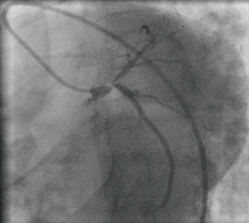
Coronary angiogram still shot showing distal left main coronary artery and tight left anterior descending coronary artery disease in a left anterior oblique caudal view. Conventionally this is an indication for coronary artery bypass grafting, though currently most operators would also consider percutaneous transluminal coronary angioplasty with stenting for this type of disease due to the availability of better PTCA hardware including drug eluting stents.
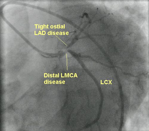
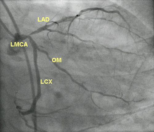
Right anterior oblique caudal view also demonstrates the ostial disease of LAD nicely. Tapering of the distal left main is also evident in this view. The circumflex ostium seems to relatively spared in this view. LCX and hence the left coronary artery is a dominant vessel here as it gives rise to the posterior left ventricular and posterior descending coronary artery. PLV and LPDA are not marked in the image, but are the terminal branches of the LCX seen at the bottom of the image.
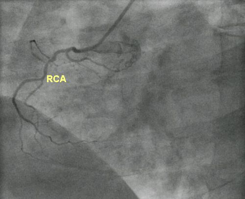
Non dominant right coronary artery is seen to be an insignificant vessel. This emphasises the urgent need to revascularise the left coronary artery territory at the earliest.
Related Posts


About The Author
Johnson Francis
Former Professor of Cardiology, Calicut Govt. Medical Kozhikode, Kerala, India. Editor-in-Chief, BMH Medical Journal