Carbon dioxide angiography
Carbon dioxide angiography

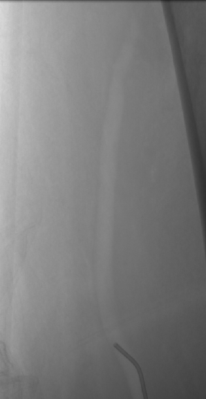
Carbon dioxide angiography (CO2 angiography) is resorted to in cases of renal failure. Iodinated contrast has the risk of contrast induced acute kidney injury, more so in those with preexisting renal insufficiency. Earlier those with renal issues were considered for magnetic resonance angiography which was also called dyeless angiography as blood vessels can be visualized with the contrast obtained from moving hydrogen nuclei in water, an important constituent of blood. But for better visualization, gadolinium based contrasts are often used in magnetic resonance angiography. Gadolinium based contrasts have been associated with nephrogenic systemic sclerosis in those with renal failure. Hence the role of carbon dioxide angiography in those with renal failure as carbon dioxide is devoid of any such toxicity. As CO2 is highly soluble in blood and rapidly cleared from circulation, it is ideally suited as a negative contrast agent in those with renal failure.
Technique of CO2 angiography is a little more cumbersome than contrast angiography. We need a medical grade carbon dioxide cylinder, three way stop cocks, connector tubings, syringes and a reservoir bag for interim storage of carbon dioxide. Initially the reservoir bag is connected to the CO2 cylinder and CO2 drawn into it multiple times to flush out the room air present in it. After that the bag can be connected to the angiographic manifold attached to the proximal end of the catheter for CO2 injection [1]. High flow rates can cause mesenteric ischemia due to vapour lock, which can be managed by rotating the patient side to side and gently massaging the abdomen.
Carbon dioxide angiography is an alternative in those with contrast allergy as carbon dioxide is non-allergenic [2]. Carbon dioxide angiography should not be used for cerebral vessels and injections should be below the level of the diaphragm. That would mean that it cannot be used in the thoracic aorta or for coronary angiography. Many of the complications of carbon dioxide angiography are at least partly contributed to by contamination with room air [3].
Occasional reports of fatality following carbon dioxide angiography prompted a study on the safety. A retrospective study of 951 patients who underwent 1007 carbon dioxide digital subtraction angiography (DSA) procedures over a 21 year period has been reported [3]. 632 were arterial procedures, which included 527 aortograms, 100 extremity arteriograms and 5 pulmonary arteriograms. 187 inferior vena cavagrams, 182 hepatic or visceral venograms, 5 extremity venograms and 1 superior vena cavagram were the venous procedures. Associated endovascular procedures were done in 499 cases. Of the four deaths in the series (0.4%), one was due to suppurative pancreatitis following aortogram and another with hepatic bleed following failed transjugular intrahepatic portosystemic shunt. Another was due to metastatic adenocarcinoma and one due to refractory end-stage cardiomyopathy.
A randomized study has been conducted on quality and safety of automated carbon dioxide DSA in femoropopliteal lesions [4]. All the fifty patients in the study underwent both iodinated contrast media and carbon dioxide angiography for the same target lesion. Inter rater agreement was fair to excellent for overall image quality. It was fair to excellent for visibility of collaterals and poor to excellent for assessment of stenoses/occlusion. Adverse events noted were hematoma in two patients, pain related to puncture in one and nausea and vomiting in another.
Indications for carbon dioxide angiography
- Abdominal aortography
- Renal transplant arteriography
- Visualization of portal vein with wedged hepatic venous injection
- For guiding endovascular interventions
References
- Funaki B. Carbon Dioxide Angiography. Semin Intervent Radiol.2008; 25: 65-70.
- Cho KJ. Carbon Dioxide Angiography: Scientific Principles and Practice. Vasc Specialist Int. 2015 Sep;31(3):67-80.
- Moos JM, Ham SW, Han SM, Lew WK, Hua HT, Hood DB, Rowe VL, Weaver FA. Safety of carbon dioxide digital subtraction angiography. Arch Surg. 2011 Dec;146(12):1428-32.
- Bürckenmeyer F, Schmidt A, Diamantis I, Lehmann T, Malouhi A, Franiel T, Zanow J, Teichgräber UKM, Aschenbach R. Image quality and safety of automated carbon dioxide digital subtraction angiography in femoropopliteal lesions: Results from a randomized single-center study. Eur J Radiol. 2021 Feb;135:109476.
